
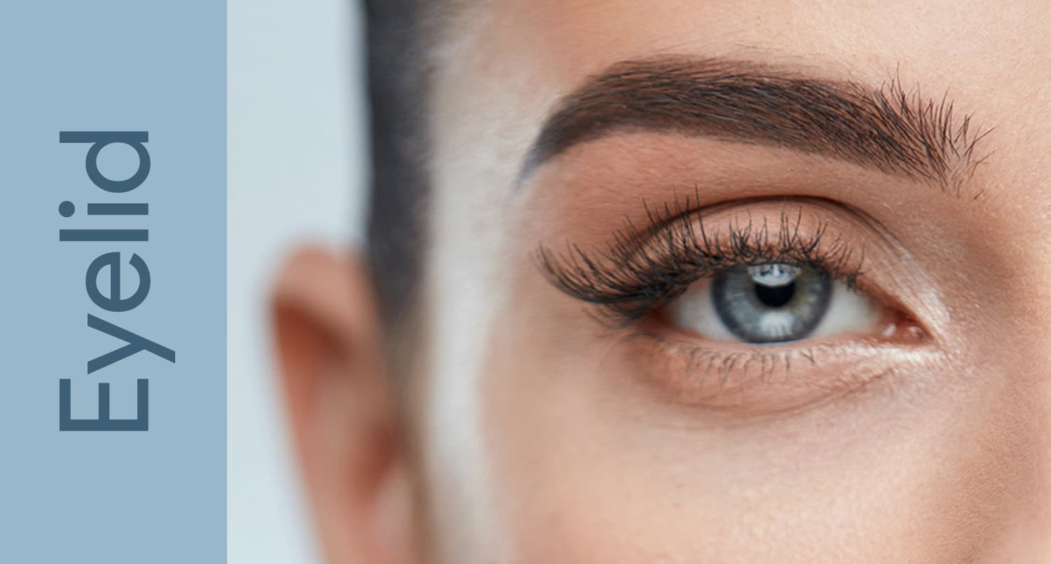
Endoscopic Brow Lift
The endoscopic brow lift uses small incisions and an endoscope to reposition the brow with minimal scarring and faster recovery.
The endoscopic brow lift is a minimally invasive technique that has largely replaced the traditional coronal brow lift for most patients seeking surgical rejuvenation of the upper face. Using a small endoscope inserted through five short scalp incisions, the oculoplastic surgeon releases the tissues that tether the brow downward and then re-suspends the brow at a more youthful position. The result is a refreshed, naturally elevated brow with minimal scarring, preserved sensation, and no visible hair loss along the incision lines.
For patients bothered by a heavy, tired-looking upper face — the kind of appearance that prompts questions like “why do you look angry?” or “are you tired?” — the endoscopic brow lift addresses the root cause rather than masking it. When combined thoughtfully with upper blepharoplasty, it produces results that other modalities simply cannot match.

Brow Ptosis Anatomy
The brow is supported by a balance of elevator and depressor muscles. The frontalis muscle — the broad muscle of the forehead — is the only true elevator of the brow. Working against it are several depressors: the corrugator supercilii (which pulls the medial brow down and inward, creating vertical frown lines), the procerus (which depresses the medial brow, creating horizontal lines at the nasal root), and the orbital portion of the orbicularis oculi (which depresses the lateral brow).
With aging, three changes converge. First, the soft tissues of the forehead lose volume and elasticity, allowing gravity to win the tug-of-war. Second, the retaining ligaments that anchor the brow to the underlying frontal bone weaken and stretch. Third, chronic activation of the depressor muscles — often unconsciously in response to a heavy brow — further pulls the brow downward. The lateral brow typically descends first and most dramatically, because the frontalis muscle has no fibers in the lateral third of the brow to oppose the orbicularis depressor action.
The endoscopic technique addresses each of these layers. By releasing the periosteum and the arcus marginalis (the fibrous attachment along the supraorbital rim), the surgeon frees the brow from its tethered position. By weakening or selectively dividing the corrugator and procerus, the depressor forces are reduced. By re-fixating the brow superiorly, the elevated position is maintained while the periosteum heals down in its new location.
For a broader overview of all surgical and non-surgical approaches to brow rejuvenation, see our main Brow Lift page, which compares endoscopic, direct, pretrichial, and temporal techniques.
Surgical Technique
The endoscopic brow lift is performed under general anesthesia or deep sedation, typically as an outpatient procedure lasting 60 to 90 minutes. The technique has been refined over the past three decades and is now highly reproducible in experienced hands.
Incision Placement
Five incisions are made behind the hairline, each measuring approximately 1 to 2 centimeters. One central incision sits at or just behind the hairline in the midline. Two paramedian incisions are placed along the brow’s ideal vector of elevation — usually about 5 to 6 centimeters from the midline. Two temporal incisions are placed obliquely behind the temporal hairline to address the lateral brow, which often requires the most elevation.
Endoscopic Dissection and Release
Through the central and paramedian incisions, the surgeon develops a subperiosteal plane — meaning dissection occurs directly on the frontal bone, beneath the periosteum. This avascular plane minimizes bleeding and protects the supratrochlear and supraorbital neurovascular bundles, which provide sensation to the forehead and scalp. The endoscope, equipped with a 30-degree angled lens, projects a magnified view onto a monitor, allowing precise identification of these critical structures.
Dissection proceeds inferiorly until the supraorbital rim is reached. The arcus marginalis — the dense fibrous attachment along the rim — is then sharply released, which is the critical maneuver that allows the brow to move upward. Laterally, the temporal dissection proceeds in the plane between the deep temporal fascia and the superficial temporal fascia. The conjoint tendon, where these layers fuse along the temporal crest, is divided to allow communication between the central and lateral dissection planes.
Depressor Muscle Modification
Once the brow is fully released, the corrugator and procerus muscles are identified and selectively weakened. The goal is not total resection — which can create a hollowed, surprised appearance — but rather thoughtful reduction of their pull. Many surgeons preserve a thin slip of corrugator to maintain natural animation.
Fixation Methods
Fixation is the step that determines longevity. Several methods are in current use:
- Cortical bone tunnels: Small tunnels are drilled into the outer cortex of the frontal bone, and sutures anchored to the periosteum are passed through these tunnels. This is considered the gold standard for durability.
- Absorbable fixation devices: Devices such as the Endotine® (a small tined absorbable implant) are placed in a drilled well in the frontal bone, and the periosteum is impaled onto the tines. The device dissolves over 6 to 12 months as scar tissue holds the new position.
- Bone screws (temporary): Titanium screws may be placed transcutaneously and removed at 10 to 14 days, allowing fibrous adhesion to mature.
- Fibrin glue: Used as an adjunct rather than primary fixation, fibrin sealant can help stabilize tissue planes during early healing.
- Temporal suture suspension: Sutures from the temporoparietal fascia to the deep temporal fascia hold the lateral brow elevation.
Advantages Over Coronal Lift
The coronal brow lift — the historical gold standard — involves a long incision extending from ear to ear across the top of the scalp. While effective, it carries significant downsides that the endoscopic approach largely eliminates.
Endoscopic Brow Lift
- Five small incisions (1–2 cm each)
- No alteration of hairline position
- Minimal hair loss along incisions
- Preserved scalp sensation in most cases
- Recovery typically 7–10 days
- Lower risk of contour irregularities
- Reduced blood loss
Coronal Brow Lift
- Long ear-to-ear incision
- Hairline often elevated 1–2 cm
- Visible scarring through hair
- Numbness behind incision common, sometimes permanent
- Recovery 2–3 weeks
- Greater scalp tension
- Greater blood loss
The endoscopic approach also preserves the option for revision. Because so little tissue is removed and the scalp is not shortened, future surgical adjustments remain straightforward. Patients with high foreheads — for whom a coronal lift would worsen the disproportion by further elevating the hairline — are particularly well-served by the endoscopic technique.
Limitations
The endoscopic brow lift is not the right operation for every patient. Understanding its limitations is essential to setting realistic expectations and choosing the best surgical plan.
Very heavy brows with significant skin excess: Because the endoscopic technique does not remove skin, patients with profound brow ptosis and abundant forehead skin redundancy may achieve insufficient elevation. In these cases, a direct brow lift (incision just above the brow hair) or a pretrichial lift (incision at the hairline that also removes forehead skin) may provide more powerful correction.
Very high hairlines: Patients with a forehead height greater than approximately 6 to 7 centimeters may notice further hairline elevation with endoscopic technique, though this is far less dramatic than with coronal lift. A pretrichial approach can actually lower the hairline in these patients.
Severe deep forehead rhytids: While the endoscopic lift softens forehead lines, it does not eliminate them. Adjunctive botulinum toxin and skin resurfacing are often needed to address etched-in lines.
Asymmetric brow ptosis from nerve injury: Patients with unilateral frontalis paralysis from prior Bell’s palsy or facial nerve injury may need a direct unilateral brow lift for symmetric correction.
Important: Patients who have unconsciously been recruiting their frontalis muscle to lift heavy brows may notice, after surgery, that their upper eyelid hooding looks worse than expected. This is because the frontalis can finally relax, revealing the true degree of eyelid skin excess. A combined plan addressing both brow and eyelid is often necessary.
Recovery
Recovery from endoscopic brow lift is substantially faster than from coronal lift, but it is not instantaneous. Understanding the typical timeline helps patients plan time off and social commitments realistically.
First 48 hours: A soft head wrap is typically worn to minimize swelling. Patients are encouraged to keep the head elevated, use cold compresses around (not directly on) the incisions, and avoid bending or straining. Mild headache and a sensation of scalp tightness are normal.
Days 3–7: Swelling peaks at 48 to 72 hours and then steadily declines. Bruising, if present, generally migrates downward into the upper eyelids and cheeks. Sutures or staples in the scalp are removed around day 7 to 10. Hair washing is permitted within 48 to 72 hours.
Weeks 2–3: Most patients are comfortable returning to work and social activities. Residual swelling is usually subtle and easily concealed. Light exercise (walking) is encouraged; aerobic exercise can resume around 2 to 3 weeks.
Weeks 4–6: Full exercise, including weight-lifting and contact sports, is permitted. Numbness or altered sensation behind the incisions gradually improves; most patients regain normal sensation by 3 to 6 months, though small areas of permanent numbness behind incision sites are possible.
Months 3–6: Final position settles as deep tissues fully heal and any absorbable fixation device dissolves. Subtle drop from the immediate post-operative position is expected and accounted for during surgery by slightly over-correcting.
Results and Longevity
Properly performed, an endoscopic brow lift delivers durable, natural-appearing results. The brow sits at a more youthful position — just at or slightly above the supraorbital rim in women, and at the rim in men — with a soft lateral arch. The forehead appears smoother, the eyes more open, and the resting expression more relaxed.
Longevity depends on several factors: the quality of fixation, the patient’s tissue elasticity, sun exposure habits, weight stability, and ongoing use of neuromodulators to suppress depressor muscle activity. Most patients enjoy meaningful improvement for 7 to 10 years, with many maintaining benefit even longer. The brow continues to age after surgery, but it ages from a more elevated starting point.

Regular maintenance with botulinum toxin to the corrugators and procerus extends results significantly by preventing the depressor muscles from gradually pulling the brow back down. Many surgeons recommend resuming neuromodulator treatments approximately 3 months postoperatively as part of the long-term maintenance plan.
Combination With Upper Blepharoplasty
One of the most important concepts in periocular rejuvenation is the relationship between the brow and the upper eyelid. The two structures are anatomically and functionally inseparable — the brow forms the upper boundary of what the patient perceives as “eyelid heaviness.” Failure to recognize and address brow ptosis when performing upper blepharoplasty is one of the most common causes of unsatisfactory outcomes.
Why Sequence Matters
When brow ptosis and dermatochalasis (excess eyelid skin) coexist, the brow lift should be performed first — or simultaneously, with the brow lift planned and marked first. Here is why:
- Brow elevation pulls eyelid skin upward and recruits it back toward the brow. The amount of redundant upper eyelid skin that needs to be removed during blepharoplasty is therefore reduced.
- If blepharoplasty is performed first and removes a generous amount of skin, a subsequent brow lift may pull the brow into the eyelid skin defect — potentially causing lagophthalmos (inability to fully close the eyes), which is a serious complication.
- Performing brow lift first protects against creating a permanently surprised or hollowed upper eyelid appearance.
For a deeper comparison of how these two procedures differ in indication and effect, see our guide on Blepharoplasty and how it relates to brow lift decision-making.
The “Brow Position Test”
Before surgery, the oculoplastic surgeon performs a simple but critical maneuver: manually elevating the brow to its anticipated post-lift position and then re-assessing the upper eyelid. If most of the apparent eyelid hooding disappears with brow elevation, the patient primarily needs a brow lift. If significant eyelid skin redundancy remains even with the brow elevated, both procedures are indicated. If only mild redundancy remains, a conservative blepharoplasty can be planned to complement the brow lift.
| Clinical Finding | Recommended Approach |
|---|---|
| Brow at or below supraorbital rim, hooding resolves with manual brow elevation | Endoscopic brow lift alone |
| Mild brow ptosis with prominent eyelid skin excess | Upper blepharoplasty alone, possibly with botulinum toxin chemical brow lift |
| Significant brow ptosis plus persistent eyelid skin excess | Combined endoscopic brow lift and upper blepharoplasty (brow first) |
| Lateral brow ptosis only | Lateral-only endoscopic or temporal brow lift |
Who Is a Good Candidate
The ideal candidate for endoscopic brow lift shares several features:
- Brow ptosis at or below the supraorbital rim, particularly with lateral brow descent creating a tired or angry appearance.
- Forehead height of 5 to 6 centimeters — not so high that further elevation would be cosmetically unfavorable.
- Adequate scalp mobility assessed during the consultation.
- Realistic expectations — understanding that brow lift creates a refreshed, natural appearance, not a dramatic transformation.
- Good general health with controlled blood pressure, no active smoking, and no medical conditions that impair healing.
- Stable weight — significant fluctuations can compromise long-term results.
Less ideal candidates include patients with very high hairlines (who may benefit from pretrichial approach), profound skin excess (who may need direct brow lift), active smokers (who heal poorly), or those whose primary concern is forehead wrinkles rather than brow position (who may do better with botulinum toxin and skin resurfacing alone).
For patients evaluating whether to pursue brow lift versus other facial rejuvenation, an honest comparison of options — including how brow lift differs from a full facelift evaluation — is essential. The brow lift addresses the upper third of the face only; it does not affect the midface, jowls, or neck. Patients with concerns spanning multiple facial zones may benefit from a staged or combined surgical plan.
Important: Choose a surgeon with specific endoscopic brow lift experience. The technique requires comfort with endoscopic visualization, familiarity with multiple fixation methods, and the ability to make intraoperative adjustments. ASOPRS-trained oculoplastic surgeons are uniquely qualified because of their deep expertise in periorbital anatomy and their ability to integrate brow surgery with eyelid surgery in a single, harmonious plan.
If you are considering brow rejuvenation and want to understand whether the endoscopic approach is right for you, the next step is a consultation with an ASOPRS fellowship-trained oculoplastic surgeon. They will evaluate your brow position, eyelid anatomy, hairline, and skin quality, and develop a personalized plan that may include endoscopic brow lift alone or in combination with other procedures. Find a Doctor in your area to schedule a consultation and learn what results are realistic for your anatomy and goals.
Frequently Asked Questions
- Who is a good candidate for an endoscopic brow lift?
- Ideal candidates are patients with mild to moderate brow ptosis (drooping) who want to restore a more youthful appearance without extensive surgery. Good candidates typically have realistic expectations, are in overall good health, and have sufficient hair coverage to conceal small incisions. Your oculoplastic surgeon will evaluate your facial anatomy and skin quality during a consultation to determine if you're a suitable candidate for this procedure.
- What should I expect during my initial consultation?
- During your consultation, your surgeon will examine your brow position, skin elasticity, and facial structure to assess your candidacy and discuss your aesthetic goals. They will explain the endoscopic technique, review before-and-after photos, and answer your questions about risks and recovery. Your surgeon may also take photographs to document your baseline appearance and help plan your personalized surgical approach.
- How does the endoscopic technique differ from traditional brow lift surgery?
- The endoscopic approach uses a small camera (endoscope) inserted through tiny incisions to visualize and lift the brow tissue, whereas traditional methods require longer incisions across the scalp. This minimally invasive technique results in less tissue trauma, reduced scarring, and typically faster healing with minimal hair loss. The small incisions are typically hidden within the hairline, making them virtually undetectable once healed.
- What are the potential risks and complications of endoscopic brow lift?
- While generally safe, potential risks include temporary numbness, nerve injury causing forehead weakness, asymmetry, and brow under-correction or over-correction. Infection, bleeding, and adverse reactions to anesthesia are possible but rare with a skilled surgeon. Most complications are minor and resolve within weeks to months, though some effects like numbness may persist longer.
- How long do the results of an endoscopic brow lift last?
- Results typically last 10-15 years, though this varies based on individual aging, skin quality, and lifestyle factors. The procedure addresses the current position of the brow but does not stop the natural aging process, so some gradual descent may occur over time. Many patients find that even after results fade, their brows remain in a better position than before surgery.
- What does post-operative care involve after my endoscopic brow lift?
- Most patients can return home the same day and resume light activities within one to two weeks, with full recovery typically occurring in four to six weeks. You'll need to avoid strenuous exercise, heavy lifting, and bending over during initial healing to prevent complications. Your surgeon will provide specific instructions on wound care, activity restrictions, and when to schedule follow-up appointments.
- When should I see an oculoplastic surgeon versus a general plastic surgeon for a brow lift?
- Oculoplastic surgeons (fellowship-trained in eye and surrounding tissue surgery) have specialized expertise in the delicate structures around the eyes and brow, which is critical for achieving natural, balanced results. If you have concerns about drooping eyelids, tear duct issues, or complex facial anatomy, an oculoplastic surgeon's focused training in these areas is particularly valuable. ASOPRS fellowship-trained surgeons have completed rigorous additional training specifically in this surgical region.
Find a Specialist
Connect with a board-certified oculoplastic surgeon who specializes in endoscopic brow lift.
Search the Directory →